
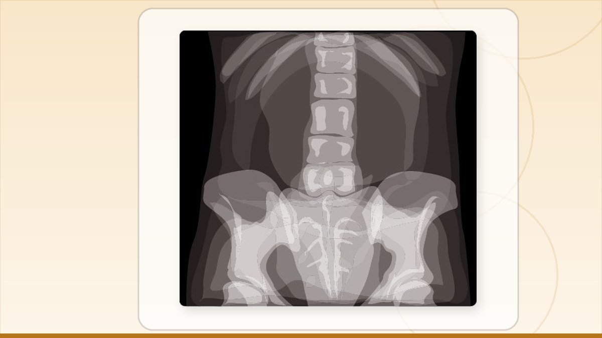
For most back pain, no scan is needed in the first six weeks. An X-ray shows bone: fractures, alignment, wear. An MRI shows discs, nerves and the spinal cord, so it is the scan that decides disc surgery. A CT shows fine bone detail when an X-ray is not enough. Your symptoms choose the scan, not the severity of pain.
What does each scan actually show?
Think of it as three different questions about your spine. An X-ray asks about bone. An MRI asks about the soft things inside: discs, nerves, the cord. A CT asks about bone again, but in far more detail than an X-ray can give.
An X-ray takes seconds, costs the least and is widely available in Faisalabad. It will show a fracture, a bone slipping forward over the one below it, narrowed disc spaces, arthritis, scoliosis. What it will never show is a slip disc pressing on a nerve. Many patients arrive holding an X-ray film that says "disc space reduced" and believe this is proof of a disc problem needing surgery. It is not. It is a shadow where a disc used to be thicker.
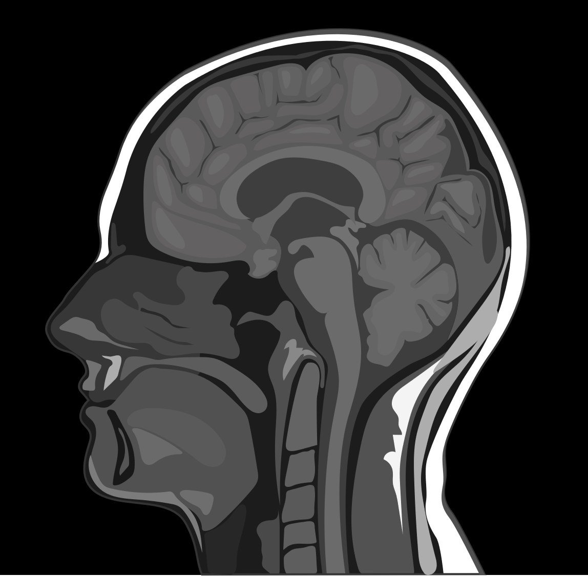
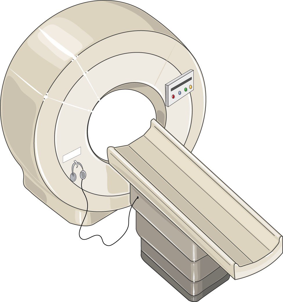
An MRI uses a magnet, not radiation. It takes twenty to forty minutes and you lie still inside a tube. It shows the disc itself, whether the disc material has come out of place, and whether it is touching or squeezing a nerve root. If a surgeon is going to operate on a disc, the operation is planned on the MRI.
Simple rule: pain only in the back, no leg symptoms, no warning signs, and it started recently, then you likely need no scan. Pain shooting down the leg or arm, numbness, or weakness that is not settling, then an MRI is the scan that answers the question.
My report says slip disc. Am I going to be paralysed?
Almost certainly not. A disc bulge on an MRI report is not a prediction of paralysis, and for the large majority of people it is not even a reason to book surgery.
Here is something most patients are never told. If you took a hundred people walking around Faisalabad with no back pain at all and put them through an MRI, a large share of them would show disc bulges, disc dehydration and degeneration. The proportion climbs steadily with age. These findings are so common that on their own they mean very little. What gives an MRI meaning is whether the picture matches your symptoms: the nerve that looks squeezed on the film must be the same nerve that supplies the exact part of your leg that hurts or feels numb.
Paralysis from a lumbar disc is rare. The situation that genuinely threatens the nerves is cauda equina syndrome, and it announces itself loudly: numbness in the saddle area between the legs, loss of control over urine or stool, and weakness in both legs. That is an emergency and needs an MRI the same day. Progressive, worsening weakness in one foot, for example a foot that starts dragging, also needs urgent assessment.
Outside those situations, take a breath. Getting the MRI does not commit you to an operation. It gives you and your surgeon information, and quite often the information supports waiting.
Do you need a scan at all in the first few weeks?
Usually not. Ordinary kamar dard, without leg pain and without warning signs, does not need imaging in the first six weeks. Mainstream guidelines everywhere say the same thing, and they say it because early scanning does not make people better and often makes them worse.
Why worse? Because the report will find something. It finds something in nearly everyone. A patient who is told at week two that they have "degenerative disc disease at L4-L5 with mild bulge" starts moving less, stops lifting, becomes frightened of their own back, and the pain lasts longer than it would have. The film has done harm.
What actually helps in those first weeks: keep moving within what you can tolerate, simple painkillers your doctor approves, heat, and going back to normal activity earlier than feels comfortable. Bed rest for days on end makes recovery slower. Most episodes of back pain settle on their own, whether or not anybody scanned them.
When is a scan genuinely urgent?
Some symptoms change the answer completely. If any of the following apply, do not wait six weeks and do not go to a hakeem first. Get seen and get imaged.
- Loss of control of urine or stool, or numbness in the saddle area between the legs
- Weakness in both legs, or weakness in one leg that is getting worse day by day
- Back pain after a fall, a road accident, or any injury in someone with thin bones or older age
- Fever with back pain, or back pain in someone with diabetes, on dialysis, on steroids, or with recent infection
- A known cancer anywhere in the body, with new back pain
- Night pain that wakes you and does not ease with position, along with weight loss you did not intend
- Back pain in a person under twenty or first starting after fifty
These are the situations where imaging exists to find something serious: infection, fracture, tumour, a nerve emergency. In every one of them, MRI is usually the right test, not X-ray.
Which scan do you need for neck pain?
The same logic, moved upward. Plain gardan dard with stiffness and no arm symptoms rarely needs an MRI early. Pain running down the arm, tingling in the fingers, or weakness of grip is the point where an MRI becomes worth doing.
There is one extra thing to watch for in the neck that has no equivalent in the lower back. The spinal cord itself runs through the neck, and pressure on the cord causes a different pattern: clumsy hands, dropping things, buttons and keys becoming difficult, unsteady walking, a feeling that the legs are heavy. If those appear, an MRI of the cervical spine should be done without delay. Cord compression treated early does much better than cord compression treated late.
For everything else in the neck, the honest best treatment for neck pain is unglamorous: posture correction, especially for people who spend hours on a phone or over a laptop, targeted physiotherapy, a proper pillow that keeps the neck neutral, and time. Most neck pain settles. Traction and long courses of massage help some people and harm others, and neither of them changes what an MRI shows.
Where does a CT scan fit in?
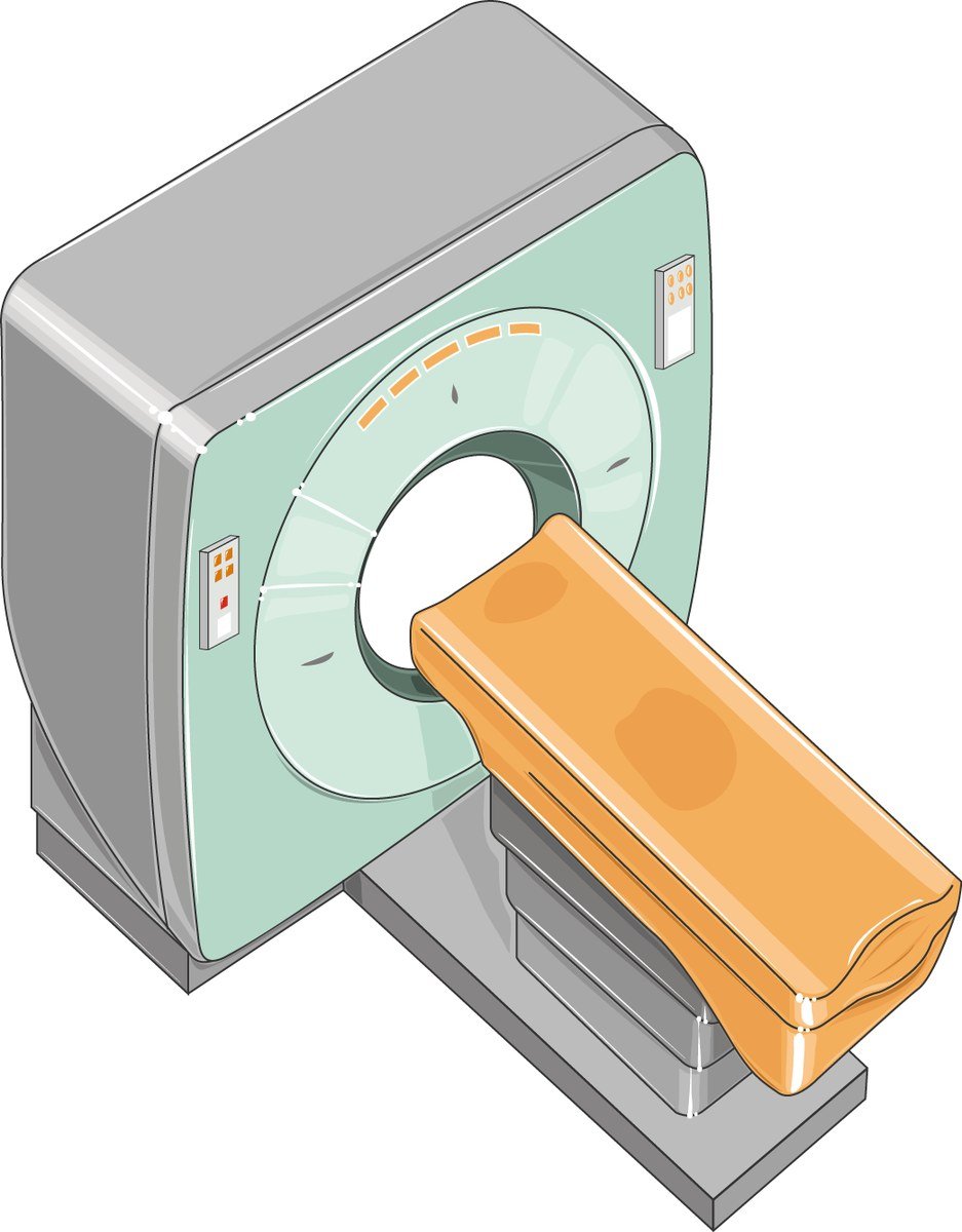
CT is a bone test. It is used when the question is about bone and an X-ray is not detailed enough, or when an MRI cannot be done.
Common reasons a spine surgeon asks for CT: a suspected fracture that the X-ray does not settle, planning of screws and instrumentation before a fusion, assessment of bone spurs or a bony canal that has become narrow, and checking how a fusion has healed afterwards. CT is also the fallback when a patient has a pacemaker, certain metal implants, or genuine claustrophobia that makes MRI impossible.
Two things to keep in mind. CT uses a meaningful dose of radiation, more than an X-ray, so it should be requested for a reason, not out of habit. And CT is poor at showing a disc pressing on a nerve. If the question is nerve compression, CT is not the answer, MRI is.
Why do so many scans in Punjab get wasted?
Because the order of events is often wrong. A very common path looks like this: months of massage and hakeem treatment first, then an X-ray from a small lab, then a second X-ray somewhere else, then an MRI on a low-strength machine with a report written in a hurry, then a second MRI at the hospital because the first films were not usable for planning. Money spent, months lost, and the disc has been pressing on the nerve the whole time.
A few things that save money and time:
- Get assessed before you get scanned. A proper examination decides which scan, or whether any scan is needed. Buying a scan first and looking for a doctor afterwards is the expensive route.
- MRI quality matters. Ask the strength of the machine. A 1.5 Tesla or 3 Tesla scanner gives images a surgeon can plan on. Very low-field machines often produce films that have to be repeated.
- Bring the images, not only the report. Ask the lab for a CD or the digital files. The report is one radiologist's summary. Your surgeon needs to look at the pictures.
- Do not repeat an MRI that is still current. If your symptoms have not changed, a scan from a few months ago is usually still valid.
Massage and traditional remedies are not evil, and for simple muscular kamar dard they sometimes give relief. The problem is delay. Forceful manipulation of a spine with an unstable fracture, an infection, or a badly compressed nerve can do real damage. If you have any of the warning signs listed above, that is not the time for malish.
The MRI shows a disc pressing on a nerve. What are the treatment options?
Even then, surgery is not automatic. A good number of disc herniations with taang mein dard improve over weeks with medication, physiotherapy and time, and the disc material itself often shrinks on its own. The first plan for most people is non-surgical treatment given a fair trial.
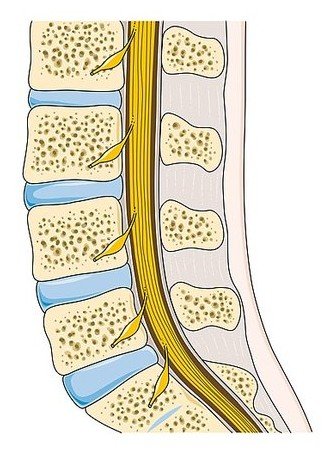
Surgery moves up the list when there is progressive weakness, when cauda equina signs appear, or when leg pain that matches the MRI has not settled after a proper course of conservative treatment and is stopping you living your life. Surgery for a disc is done to relieve the leg pain and free the nerve. It is less reliable for back pain alone, and any surgeon who promises otherwise is overselling.
Two operations do most of this work:
- Endoscopic spine surgery. A small camera and instruments are passed through an incision roughly the width of a fingertip. The muscle is spread rather than cut, blood loss is small, and most patients are up and walking the same day with a short hospital stay.
- Microdiscectomy. A slightly larger opening with an operating microscope. It is a long-established, well-studied operation and remains the right choice for certain disc positions and certain anatomy.
Which one suits you depends on where the disc fragment sits, whether the bony canal is narrowed, and your own anatomy. That decision comes off the MRI, which is why the quality of the MRI matters so much.
On cost: an MRI of one region of the spine costs a small fraction of what any operation costs, and prices vary widely between labs in Faisalabad and Lahore, so it is worth asking two or three. For back surgery cost in Pakistan, never accept a verbal figure. Ask for a written quote in rupees that separates the surgeon's fee, anaesthesia, implants if any, operation theatre, room category and the expected number of nights. Ask what happens to the price if you stay longer. Bring a family member to that conversation, because these decisions are made as a family and the person paying should hear the numbers directly.
What should you do next?
Match your situation to one of these three.
Back or neck pain for less than six weeks, no leg or arm symptoms, no warning signs. You do not need a scan yet. Keep moving, take simple pain relief, avoid long bed rest, and see your doctor if it is not improving by six weeks.
Pain running down the leg or arm, numbness or tingling that is not settling. See a spine specialist and let the examination decide the imaging. If an MRI is indicated, get it on a good machine and keep the digital files.
Any warning sign: bladder or bowel change, saddle numbness, weakness getting worse, fever, injury, cancer history. Go to a hospital today. This is not a wait and watch situation.
If you already have films and reports and you do not know what they mean, bring them to a consultation and have them explained properly, including the option of not operating. Dr. M. Abdur Rehman is a consultant neurosurgeon and spine surgeon at Mujahid Hospital, Faisalabad. Patients across Punjab searching for the best endoscopic spine surgeon in Faisalabad are usually looking for the same thing: a straight answer about whether they need an operation. Bring your MRI images on a CD, bring a family member, and ask directly what happens if you choose not to have surgery. A surgeon who answers that question clearly is one you can trust with the answer to the other one.
Cervical spine surgery in Faisalabad Read the full page on cervical spine surgery in faisalabad, including who it suits, what recovery looks like and how to arrange a review.
Questions patients ask about this
Is MRI better than X-ray for back pain?
Better for a different question. X-ray answers questions about bone: fractures, alignment, slippage. MRI answers questions about discs, nerves and the spinal cord. If your problem is leg pain from a suspected slip disc, MRI is the scan that matters. For a suspected fracture, X-ray comes first.
Can an X-ray show a slipped disc?
No. An X-ray cannot image disc material or nerves. It may show a narrowed disc space, which suggests the disc has thinned with age, but it cannot show whether disc material has come out and is pressing on a nerve. Only an MRI answers that question.
Is MRI harmful or does it use radiation?
An MRI uses a strong magnet and radio waves, not radiation, and can be repeated safely. Tell the staff beforehand about a pacemaker, cochlear implant, metal in the eye, or any implanted device. The main difficulties are lying still for half an hour and the noise of the machine.
How long should I try physiotherapy before considering surgery?
For leg pain from a disc, a fair trial is usually around six to twelve weeks of proper treatment, not a few sessions. The clock stops early if weakness is getting worse, or if bladder or bowel control changes, since those need urgent assessment rather than more physiotherapy.
Do I need a scan before I see a spine surgeon?
No, and it often saves money to be examined first. The examination decides which scan is needed, and sometimes decides that none is. If you already have films, bring the digital images on a CD rather than only the printed report, since your surgeon needs to see the pictures.
If surgery is on the table
Once you have the MRI report, these explain what the findings mean for treatment.
Related articles
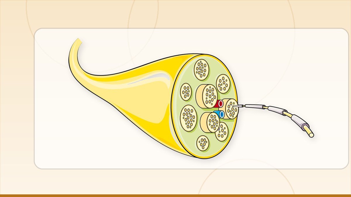
Nerve tests (EMG and NCS) for back and leg pain
Does an EMG test for sciatica tell you if you need surgery? What EMG and NCS show, when they help, what they…
Read this
Kamar dard: causes, and when it is more than muscle pain
Kamar dard ka ilaj: what causes back pain, what to try at home first, when an MRI is needed, and the small…
Read this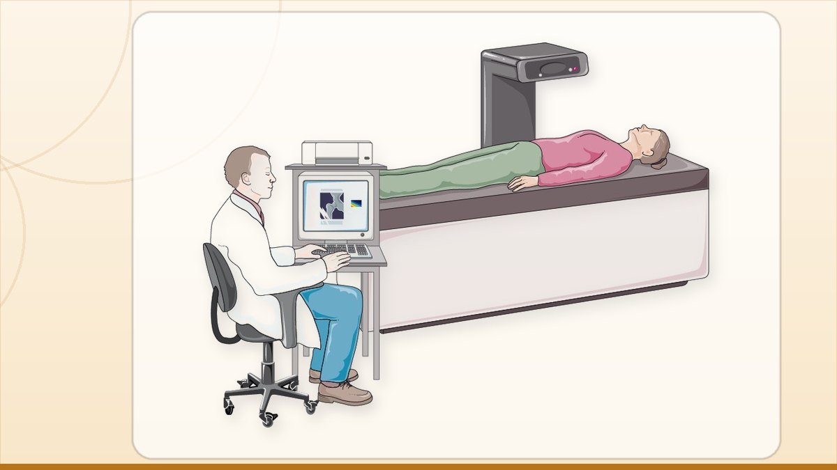
Taang mein dard: leg pain from the back, explained simply
Taang mein dard ka ilaj: leg pain usually starts from a nerve in the lower back. How to tell nerve pain from…
Read thisAll patient questions · Spine surgery in Faisalabad · Book an appointment
Ask about your own scan. Send your MRI or X-ray on WhatsApp and get a straight answer on whether surgery is needed at all, from a consultant neurosurgeon in Faisalabad.
Dr. M. Abdur Rehman is a consultant neurosurgeon in Faisalabad. Send your scan on WhatsApp and ask what it actually shows.
Published for the practice of Dr. M. Abdur Rehman, consultant neurosurgeon, Mujahid Hospital, Faisalabad. General information only, not a substitute for examination.